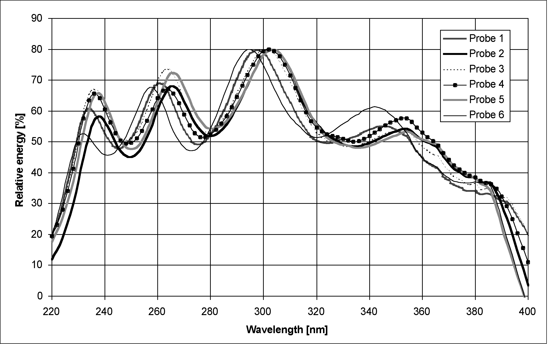
The plot of relative probe energy in artificial intestinal fluid pH 7.5 (Figure 1) shows values exceeding 30% from about 230 to 390 nm. Hence this wavelength range is suitable for measuring UV absorbance.
Evaluation of the Rainbow Dynamic Dissolution Monitor™ Semi-automatic Fiber Optic Dissolution Tester
Caspar Schatz, Michel
Ulmschneider, Rolf Altermatt, Stephan Marrer
Pharmaceutical Quality
Assurance and Quality Control, F. Hoffmann-La Roche Ltd, Basel,
Switzerland
email:caspar.schatz@roche.com
Summary
The Rainbow Dynamic Dissolution Monitor™ (Delphian Technology
LP, Ardsley, USA) is a simple and convenient UV absorbance technique
for acquiring precise, accurate, reproducible and robust dissolution
profiles of drug formulations containing a single active ingredient.
The instrument and its software are GMP compliant. Benefit analysis
shows that it has significant advantages for dissolution over
systems using filtering and flow -through cells. The Rainbow Dynamic
Dissolution Monitor™ is thus suitable for routine dissolution
analysis in pharmaceutical quality control.
Introduction
The Rainbow Dynamic Dissolution Monitor™
uses 12 fiber optic immersion probes residing in vessels of two
dissolution baths throughout the dissolution test. Two deuterium
lamps are used as a light source. After interacting with the sample,
the light is guided to a series of 12 photo diode array ultraviolet
monolithic miniature spectrometers (UV MMS, Carl Zeiss, Jena,
Germany) [1] which measure from 200 to 400 nm with an absolute
wavelength accuracy of 0.2 nm and a temperature drift less than
0.005 nm/K. The spectral pixel spacing is 0.8 nm, giving a Rayleigh
resolution of about 3 nm. Stray light measured at 240 nm using
a deuterium lamp and potassium iodide solution (10 g/l) is 0.3
% [2]. Each spectrometer unit and its probe are referred to as
a channel.
Before performing a dissolution run
the system collects 0% and 100 % transmission blanks and standard
absorbance scans per channel. Hence the amount of dissolved active
compound is determined with a single point calibration. To eliminate
standard preparation errors, a second quality control standard
is measured for control purposes only. During a run, the system
can acquire absorbance scans every 10 seconds. The software incorporates
two methods to correct for turbidity and scattering effects. The
first method uses two wavelengths: one to determine the active
compound, the other to act as a compensation wavelength. The second
method is based on a second derivative algorithm using a wavelength
range [1]. It uses a very simple algorithm to estimate the second
derivative and a form of co-addition of several wavelengths to
improve the signal to noise ratio.
Experimental
All the experiments were performed using the Rainbow Dynamic Dissolution
Monitor™ with 10 mm pathlength Hellma ultra mini-immersion
probes, (type 661.673-UV, Hellma GmbH & Co., Müllheim/Baden,
Germany) to acquire UV measurements. Only six channels/probes
were evaluated, always using one scan per measurement in each
case.
System
suitability
Suitability was assessed in terms of fiber optic unit transmission
and the linear range of the spectroscopic assembly.
Transmission
The relative energy of 100% transmission spectra of artificial
intestinal fluid pH 7.5 (reference Anticoagulant Tablets Section,
page 10, for fluid composition) was plotted against wavelength
as a measure of channel and probe transmission
Linear
range
The linear range of all six evaluated channels was tested using
a dilution series of potassium dichromate (spectroscopy grade,
Fluka Chemie AG, Buchs, Switzerland) in 0.01 N sulphuric acid
[3]. A stock solution was diluted to concentrations giving absorbance
values ranging from 0.2 to 2.0. Based on the absorbance spectrum
of potassium dichromate in 0.01 N sulphuric acid, absorbance was
measured at 258 nm.
A correlation coefficient was calculated using the absorbance
values from the two weakest standard solutions; the same process
was repeated for increasing strengths of standard solutions to
determine the upper end of absorbance linearity (taken as 99.9%
correlation with prediction).
Anticoagulant
tablets
The Rainbow Dynamic Dissolution Monitor™ was evaluated using
anticoagulant tablets containing 3 mg of active compound, corn
starch white, lactose powder, magnesium stearate, and talc.
In routine dissolution analysis,
the anticoagulant tablets are dissolved in 900 ml of artificial
intestinal fluid pH 7.5 (comprised of 80.5g anhydrous dipotassium
hydrogen phosphate and 15.6 g of potassium dihydrogen phosphate
dihydrate in 10 liters ofdistilled water), stirred at 50 rpm in
apparatus 2 [4]. The medium is degassed and heated to 37.0 ±
0.5 °C. The 20-minute Q value used for release analysis is
75% [5].
Linearity of absorbance
readings
To identify a suitable detection wavelength, triplicate absorbance
spectra were acquired of solutions equivalent to 25, 50, 75, 100,
and 125% of active compound dissolved in artificial intestinal
fluid pH 7.5, with approximate concentrations of 0.00083, 0.00167,
0.00250, 0.00333, and 0.00417 mg/ml, respectively. The resulting
correlation coefficient was plotted against wavelength.
Linearity
of compensation methods
Based on the absorbance and second derivative spectrum of active
compound in artificial intestinal fluid pH 7.5 determined in an
earlier experiment, the linearity of three different turbidity
compensation methods was investigated (Table 1).
Table 1: Three different turbidity compensation methods.
|
|
|
|
|
|
|
|
|
|
|
|
|
|
|
|
All methods were evaluated in triplicate
using 25, 50, 75, 100, and 125% solutions of active compound dissolved
in artificial intestinal fluid pH 7.5 with approximate concentrations
of 0.00083, 0.00167, 0.00250, 0.00333, and 0.00417 mg/ml, respectively,
and using clear medium as well as medium containing a concentration
of placebo powder equivalent to one 130.0 mg tablet dissolved
in 900 ml artificial intestinal fluid pH 7.5.
The validation of analytical methods
(VoAM) program, version 3.0 [6], was used, with the following
acceptance criteria [7, 8]: correlation coefficient > 0.99;
y intercept within the 95% confidence interval of 2% of the reference
x value (100% solution of active compound); precision, expressed
as the standard deviation of relative repeatability (treating
each set of triplicate data as one group), < 2.00% assuming
data and mean recovery between 98.00 and 102.00%.
Comparison of turbidity
compensation methods
Six absorbance readings of active compound solution in artificial
intestinal fluid pH 7.5 before and after addition of placebo powder
were acquired in triplicate to compare the accuracy and efficacy
of the three turbidity compensation methods, using 100% solutions
of active compound (approximately 0.00333 mg/ml).
The VoAM 3.0 program [6] was used
to determine statistical equivalence, with the following acceptance
criterion: the 95% confidence interval of the mean of the test
method had to lie entirely within 2.00% either side of the mean
of the reference method.
Robustness of the turbidity
compensation methods
Two absorbance measurements were acquired at 12 different positions
(hence different bending radii) of the fiber optic immersion probes
and cables to test the robustness of each compensation method
with respect to obligatory movement by the fiber optic immersion
probes during the performance of a dissolution run. This experiment
was performed using artificial intestinal fluid pH 7.5 containing
active compound at approximately 0.00417 mg/ml, equivalent to
the extent of dissolution of 125%.
Method comparison
The two turbidity compensation wavelength methods were compared
in dissolution runs using a dissolution bath (Distek Premiere
5100, Distek Inc., North Brunswick, USA) and three lots of anticoagulant
tablets (six tablets per lot). Active compound release was quantified
at 20 minutes using the two turbidity compensation methods and
the corresponding reference methods. With the reference methods,
a 20 ml aliquot was manually removed from each vessel and membrane-filtered
(0.45 mm pore size, Gelman Acrodisc, product no. 4496, Pall Gelman
Sciences, Ann Arbor, USA) [5]. Single point calibration was used
for quantification on a diode array spectrometer (HP 8452 A, Agilent
Technologies, Rockaway, USA) in combination with the same compensation
wavelength used with the Rainbow Dynamic Dissolution Monitor™.
Equivalence was defined as a maximum deviation of ± 2.0%
per tablet, with post-calculation rounding.
Results
System
suitability
Evidence for the suitability of fiber optic transmission and system
linear range is given below.
Transmission
The plot of relative probe energy in artificial intestinal fluid
pH 7.5 (Figure 1) shows values exceeding 30% from about
230 to 390 nm. Hence this wavelength range is suitable for measuring
UV absorbance.
Figure 1 Plot of relative immersion probe energy in artificial intestinal fluid pH 7.5 against wavelength (nm)
Linear range
The spectrum of potassium dichromate in 0.01 N sulphuric acid
shows an absorbance maximum at 258 nm being used to evaluate linear
range. Table 2 gives the upper limits of the linear ranges
examined with potassium dichromate in 0.01 N sulphuric acid at
258 nm with all six probes (channels).
Table 2: Upper end of linear range for all probes (channels).
|
|
|
|
|
|
|
|
|
|
|
|
|
|
|
|
|
|
|
|
|
Since probes 4 to 6 showed values
of 1.5 AU, the linear range of the whole system also had to be
set to 1.5 AU.
Anticoagulant
tablets
Linearity of the absorbance
readings
Figure 2 plots the correlation coefficient against
wavelength with active compound dissolved in artificial intestinal
fluid pH 7.5 (25125% solutions, with approximate concentrations
of 0.00083 0.00417 mg/ml). The active compound absorbance
spectrum in this figure indicates decreases in absorbance at 260
and 340 nm, arising from a decrease in system linearity owing
to a decrease in system signal to noise ratio. The further slight
decrease near 220 nm arises from the reduced energy available
in the shortwave UV region.
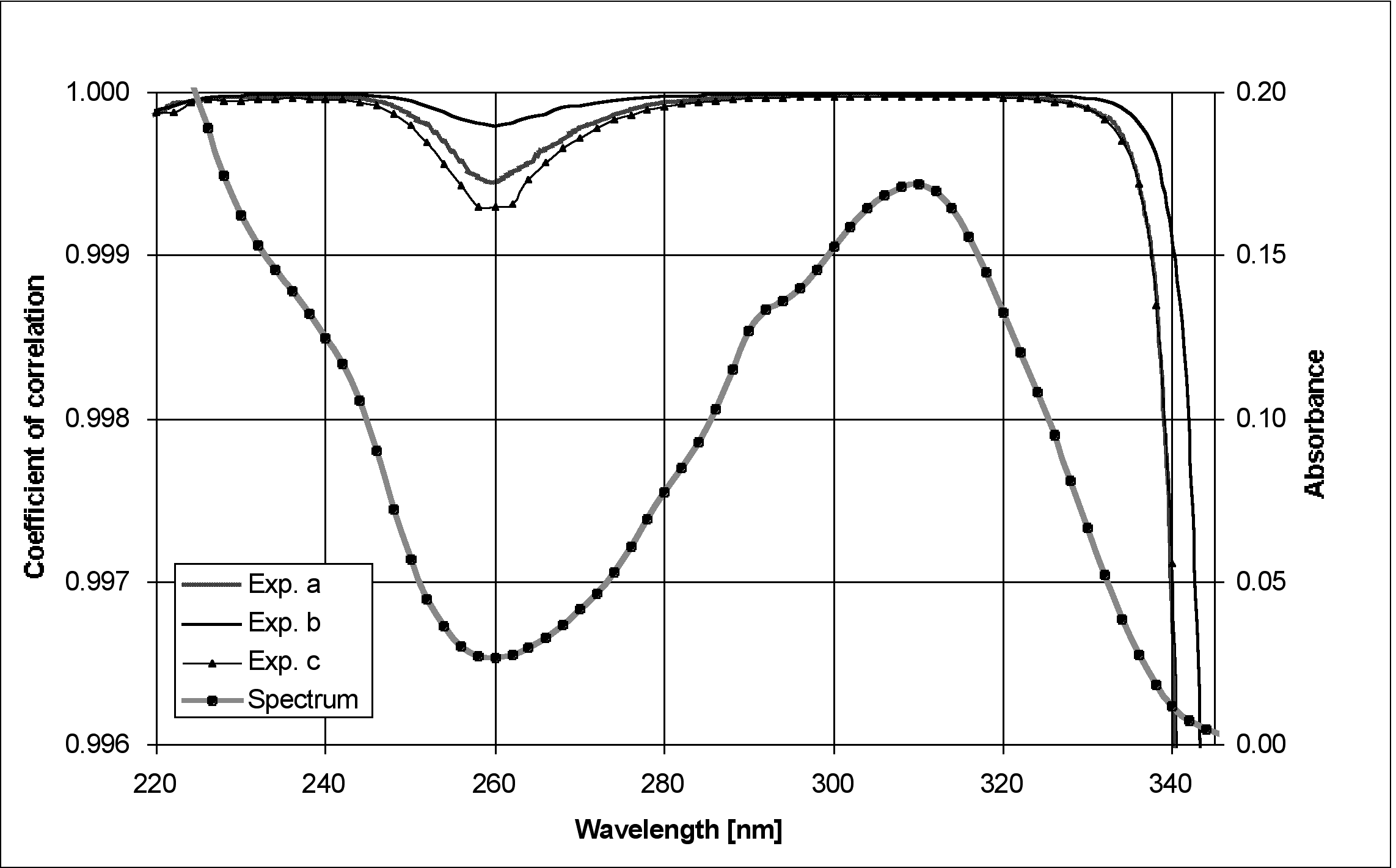
Figure 2 Linearity experiment in triplicate (ac) with active compound at a concentration equivalent to 100% dissolution in artificial intestinal fluid pH 7.5 (approximately 0.00333 mg/ml): plot of correlation coefficient against wavelength, incorporating the absorbance spectrum of active compound
It is clear that the whole wavelength
range from 220 to 340 nm is suitable for method development
Linearity of turbidity
compensation methods
Figure 3 shows the UV absorbance spectrum of active
compound and its estimate of the second derivative used in the
Rainbow Dynamic Dissolution Monitor™ software.
Based on the absorbance spectrum with a maximum at 310 nm, two
turbidity compensation methods using the peak wavelength and compensation
wavelengths of 350 and 376 nm respectively were chosen. The 300320
nm range was used in the case of the second derivative.
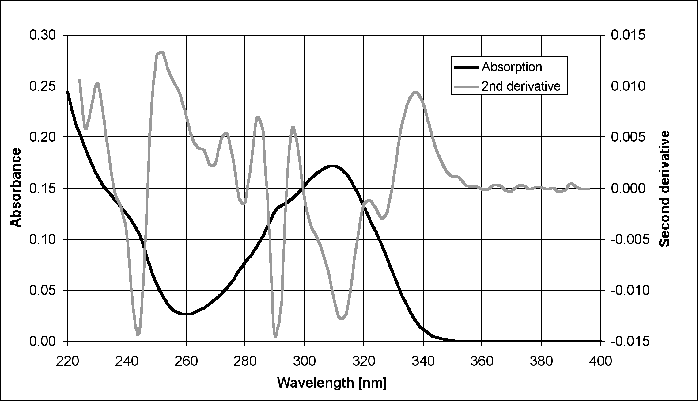
Figure 3 Absorbance spectrum of active compound at a concentration equivalent to 100% dissolution in artificial intestinal fluid pH 7.5, co-plotted with its estimate for the second derivative used in the Rainbow Dynamic Dissolution Monitor'
Table 3
gives the relevant statistical parameters for validating the three
methods using clear as well as placebo-spiked solutions.
|
|
||||||
| Parameter |
|
|
|
|
|
|
| r |
|
|
|
|
|
|
| SDrel [%] |
|
|
|
|
|
|
| intercept |
|
|
|
|
|
|
| recovery [%] |
|
|
|
|
|
|
|
|
||||||
| Parameter |
|
|
|
|
|
|
| r |
|
|
|
|
|
|
| SDrel [%] |
|
|
|
|
|
|
| intercept |
|
|
|
|
|
|
| recovery [%] |
|
|
|
|
|
|
|
|
||||||
| Parameter |
|
|
|
|
|
|
| r |
|
|
|
|
|
|
| SDrel [%] |
|
|
|
|
|
|
| intercept |
|
|
|
|
|
|
| recovery [%] |
|
|
|
|
|
|
|
|
||||||
| Parameter |
|
|
|
|
|
|
| r |
|
|
|
|
|
|
| SDrel [%] |
|
|
|
|
|
|
| intercept |
|
|
|
|
|
|
| recovery [%] |
|
|
|
|
|
|
|
|
||||||
| Parameter |
|
|
|
|
|
|
| r |
|
|
|
|
|
|
| SDrel [%] |
|
|
|
|
|
|
| intercept |
|
|
|
|
|
|
| recovery [%] |
|
|
|
|
|
|
|
|
||||||
| Parameter |
|
|
|
|
|
|
| r |
|
|
|
|
|
|
| SDrel [%] |
|
|
|
|
|
|
| intercept |
|
|
|
|
|
|
| recovery [%] |
|
|
|
|
|
|
In the case of the clear solutions
all parameters were inside the acceptance limits. There were no
significant differences (p = 95%) in method validation. However,
correlation coefficients were clearly lower, and standard deviations
of relative repeatability and recovery rates clearly higher, with
the second derivative method (Method 3) than with either compensation
wavelength method (Methods 1 and 2). Therefore it can be concluded
that the second derivative algorithm is less accurate and less
precise than either wavelength method when examining clear solutions.
When performing the same method validation
experiments with placebo-spiked solutions, there were air bubbles
in the measurement compartments of probes 1 and 4 in at least
one of the triplicate measurements. As can be seen in Figure
4, in contrast to the wavelength-independent baseline offset
caused by tablet excipients, air bubbles have a wavelength-dependent
impact on the baseline owing to the wavelength dependency of refraction
and diffraction. This explains why in Method 1, where the compensation
wavelength approximates to the analytical wavelength, only probe
4 failed the validation acceptance limits; in Method 2, on the
other hand, where the compensation wavelength is further from
the analytical wavelength, probes 1 and 4 failed the acceptance
criteria. Since the second derivative algorithm corrected for
sloping baseline offsets, all probes met the acceptance criteria,
making this the most robust method. Although there were no significant
differences (p = 95%) in method validation, the second derivative
algorithm tended to have a slightly higher standard deviation
of relative repeatability.
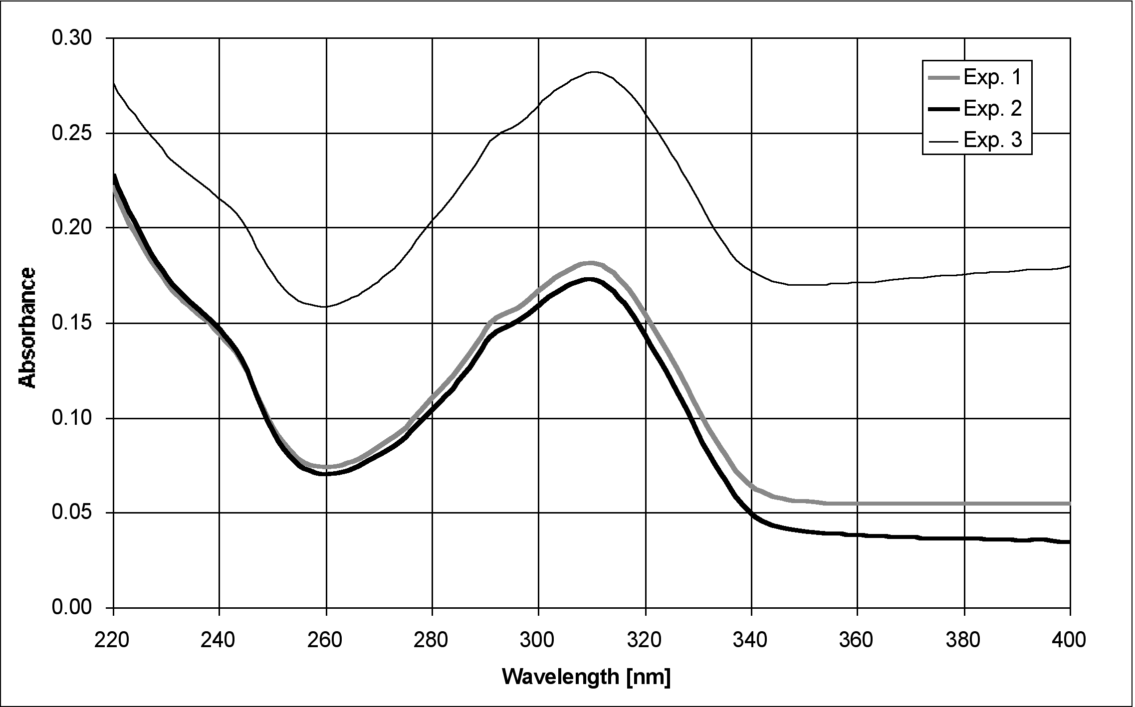
Figure 4 Probe 4: Absorbance spectra of measure 2 per triplicate (Methods 13) at a concentration equivalent to 75% dissolution (approximately 0.0025 mg/ml), comparing the wavelength-dependent baseline offset caused by air bubbles in the measuring compartment (Expts. 2 & 3) vs the air bubble-free spectrum (Expt. 1) which shows only the wavelength-independent offset caused by excipient turbidity.
Based on the validation acceptance
criteria for method equivalence there were no significant differences
(p = 95%) between the methods used for turbidity compensation.
But as can be seen from Figure 5, which shows the mean
concentrations of six measurements with their standard deviations
as error bars for the three methods before and after addition
of placebo powder, the two-wavelength compensation methods (methods
1 and 2) have a smaller standard deviation than the second derivative
method (method 3) and show less probe to probe variation. Hence
methods 1 and 2 are more rugged from this standpoint.
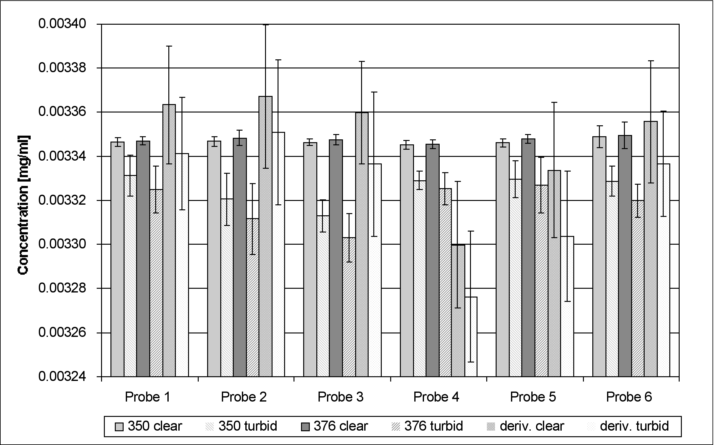
Figure 5 Concentrations measured using the three methods before and after addition of placebo powder (means of six measures). Error bars: 2 SD.
Since concentrations are higher before
than after the addition of placebo powder, all three turbidity
compensation methods overcompensate. Although the differences
are not significant (p = 95%) for method validation, the differences
between clear and turbid solutions are smallest when using the
two-wavelength method with a compensation wavelength approximating
to the analytical wavelength. Hence method 1 is most suitable
in terms of the accuracy of turbidity correction.
Robustness of turbidity compensation methods
Table 4 shows
that the method to method difference, expressed as the relative
SD, was not significant (p = 95%) in method validation. All three
methods are therefore equivalent in terms of the robustness of
moving fiber optic probes.
Table 4 Relative standard deviations (SDrel) of two measurements in 12 positions per method
|
|
||||||
|
|
|
|
|
|
|
|
| SDrel [%] |
|
|
|
|
|
|
|
|
||||||
|
|
|
|
|
|
|
|
| SDrel [%] |
|
|
|
|
|
|
|
|
||||||
|
|
|
|
|
|
|
|
| SDrel [%] |
|
|
|
|
|
|
Method comparison
Every dosage form met the acceptance criteria using method 1 and
2 (Table 5). Both methods give accurate results on the
Rainbow Dynamic Dissolution Monitor™.
Table 5 Amounts (%) of dissolved active compound using methods 1 and 2. With the Hewlett-Packard spectrometer, sample absorbances were measured after filtration. Some calculated differences do not quite match the percentages in the results column due to the calculation being performed before rounding.
|
|
|
|
||||
|
|
|
|
|
|
|
|
|
|
|
|
|
|
|
|
|
|
|
|
|
|
|
|
|
|
|
|
|
|
|
|
|
|
|
|
|
|
|
|
|
|
|
|
|
|
|
|
|
|
|
|
|
|
|
|
|
|
|
|
||||
|
|
|
|
|
|
|
|
|
|
|
|
|
|
|
|
|
|
|
|
|
|
|
|
|
|
|
|
|
|
|
|
|
|
|
|
|
|
|
|
|
|
|
|
|
|
|
|
|
|
|
|
|
|
|
|
|
|
|
|
||||
|
|
|
|
|
|
|
|
|
|
|
|
|
|
|
|
|
|
|
|
|
|
|
|
|
|
|
|
|
|
|
|
|
|
|
|
|
|
|
|
|
|
|
|
|
|
|
|
|
|
|
|
|
|
|
|
Benefit
analysis
Table 6 presents the results of a benefit analysis
comparing the Rainbow Dynamic Dissolution Monitor™ with a
conventional system using filtration and flow-through cuvettes
to determine the amount of dissolved active compound, in terms
of the following parameters: laboratory work, validation burden,
maintenance, analytical information and GMP compliance.The total
scores show that the Rainbow Dynamic Dissolution Monitor™
outperforms a semi-automatic filtering and flow-through cuvette
measurement on-line system without loss of GMP compliance.
Table 6 Benefit analysis: Rainbow Dynamic Dissolution MonitorÔ vs a conventional online system, in terms of criteria ranked using a weighting factor (0-100%). Mark (1-5): system approximation to criteria. Score: weighting factor x mark.
| Criterion |
|
|
|
||
|
|
|
|
|
||
| Laboratory work |
|
|
|
|
|
| Qualification burden |
|
|
|
|
|
| Maintenance |
|
|
|
|
|
| Analytical information |
|
|
|
|
|
| GMP compliance |
|
|
|
|
|
| Total |
|
|
|
|
|
Laboratory work
Laboratory workload, in terms of preparing the bath and standards,
is similar with both systems. During operation, no more hardware
problems are to be expected with the Rainbow Dynamic Dissolution
Monitor™ than with the conventional system since the fiber
optic immersion probes and related mechanics are quite robust
[9].
Qualification burden
Since the Rainbow Dynamic Dissolution Monitor™ incorporates
no filtration facility or liquid pump, there is less equipment
to qualify. The UV detectors are also simpler to qualify than
spectrometers used for UV/VIS precision measurements [10].
Maintenance
The Rainbow Dynamic Dissolution Monitor™ is easier to maintain
and less labor- intensive due to the elimination of sample removal
and filtration.
Analytical information
The Rainbow Dynamic Dissolution Monitor™ can supply a data
point every 10 seconds, giving dissolution profiles containing
a lot of information. It also eliminates problems due to dead
volume, time differences between sampling and measuring, and filter
clogging, leading to greater accuracy.
GMP compliance
Both systems are GMP compliant.
Conclusions
The analytical results confirm that the Rainbow Dynamic Dissolution
Monitor™ can be used to measure dissolution by methods which
meet the acceptance criteria for linearity, accuracy, precision,
and reproducibility stipulated in current validation of analytical
methods guidelines [7]. Both instrument and software are GMP compliant.
Benefit analysis shows that it outperforms dissolution measurement
systems employing filtering and flow-through cells. The advantages
have an impact on the high acquisition costs, though. The Rainbow
Dynamic Dissolution Monitor™ is thus suitable for routine
dissolution analysis in pharmaceutical quality control.
Both the two-wavelength compensation method and the second derivative algorithm are suitable for monitoring dissolution. The former is generally more precise and accurate, especially for non-disintegrating formulations where the medium stays clear. For formulations giving a background resulting in a sloping offset, the second derivative algorithm is preferable, as also when there are air bubble problems.
References
[1] Bynum K, Kraft E, Pocreva J, Ciurczak E, Palermo P, In Situ
Dissolution Testing Using a Fiber Optic Probe Dissolution System,
Dissolution Technologies, 6 (4) 810 (1999)
[2] Product information: MMS UV Monolithic Miniature Spectrometer,
Carl Zeiss, OEM Spekralsensorik, Jena
[3] European Pharmacopoeia, 3rd ed., Council of Europe, Strasbourg,
Absorbance Spectrometry, Ultraviolet and Visible, 28-29 (1997)
[4] USP 24 NF 19, United States Pharmacopeial Convention, Inc.,
Rockville MD, Dissolution, 1941-1943 (1999)
[5] Pharma Switzerland, Quality Assurance and Quality Control,
Analysis Instruction for Anticoagulant Tablets, F. Hoffmann-La
Roche Ltd, Basel (2000)
[6] VoAM 3.0, Program for Validation of Analytical Methods, F.
Hoffmann-La Roche Ltd, Basel (1999)
[7] Pharma Switzerland, Quality Assurance and Quality Control,
Guideline for Validation of Analytical Methods, F. Hoffmann-La
Roche Ltd, Basel (1998)
[8] International Conference on Harmonization, Validation of Analytical
Procedures: Methodology, ICH Harmonised Tripartite Guideline Q2B
(1995)
[9] Schatz C, Ulmschneider M, Altermatt R, Marrer S, Altorfer
H, Manual In Situ Fiber Optic Dissolution Analysis in Quality
Control, Dissolution Technologies, 7 (2) 6-13 (2000)
[10] Pharma Switzerland, Quality Assurance and Quality Control,
SOP for Testing and Maintenance of UV/VIS Precision Spectrophotometers,
F. Hoffmann-La Roche Ltd, Basel (1999)